| CEST | Tuesday, 17th September 2024 |
|---|---|
| 10:00 - 11:00 |
|
| 11:00 - 12:00 |
|
| 12:00 - 13:00 | |
| 13:00 - 14:00 |
|
| 14:00 - 15:00 |
|
| 15:00 - 15:15 | |
| 15:15 - 16:15 |
|
| 16:15 - 17:15 |
|
| 17:15 - 17:30 | |
10:00 - 11:00
Open Problems in Cardiac Ultrasound
Speaker: Andrej Varchola & Alexey Karimov (Siemens Healthineers)
Room: 335
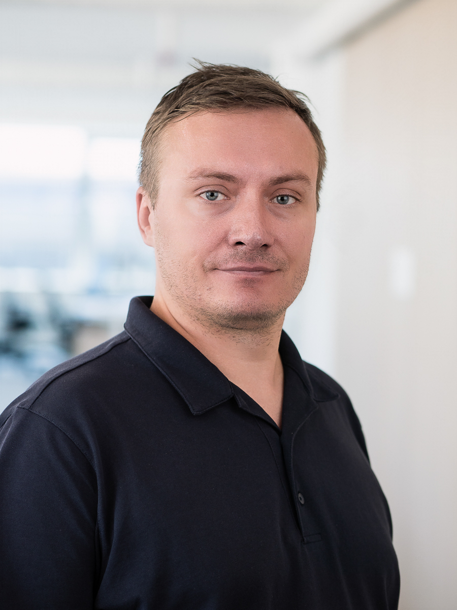
Andrej Varchola is a software engineer and technology field owner specializing in 4D visualization at Siemens Healthineers Ultrasound in Košice, Slovakia. He earned his Master's degree in Digital Signal Processing from the Slovak University of Technology and holds a PhD in Medical Visualization from the Vienna University of Technology. Andrej has successfully transitioned his academic expertise into the industry, focusing on research and development for mission-critical software. His career highlights include key contributions to several notable projects: 1) Developing HDlive rendering for realistic fetal imaging at GE Healthcare during his PhD studies. 2) Contributing to the dual-energy CT imaging system, Somatom Force, at Siemens Healthcare. 3) Visualizing data for the statistical validation of Honda's SAE Level 3 Automated Driving System. 4) Enhancing clinical applications for the Acuson Origin cardiovascular ultrasound system at Siemens Healthineers. Andrej's work bridges the gap between cutting-edge technology and practical medical applications, driving advancements in imaging and diagnostics.
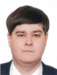
Alexey Karimov is a software engineer in the Ultrasound division of Siemens Healthineers in Vienna, Austria. He received his PhD degree from the Technical University of Vienna, working in the fields of medical visualization and real-time volume rendering. In his current position, he aims to integrate advanced rendering and visualization methods into ultrasound systems deployed in clinical environments.
Abstract: Ultrasound imaging has always been a special case in the medical domain, given that it delivers images in real-time. This combined with characteristic noise and artifacts poses a challenge to producing clinically relevant data. The newest AI algorithms come here to aid the decision process with detection, labeling, assisted navigation, and report generation. As anatomical models of heart structures are inferred and available in real time, the need for advanced visualization techniques arises. Volume rendering alone does not suffice the requirements of the field, because many more data elements are present. Seamless integration of mesh models, measurement results, and inferred anatomical labels and features are expected from the upcoming generation of rendering/visualization pipelines in ultrasound.
11:00 - 12:00
Ask-Me-Anything Session with Prof. Bernhard Preim
Speaker: Bernhard Preim (University of Magdeburg)
Room: 335
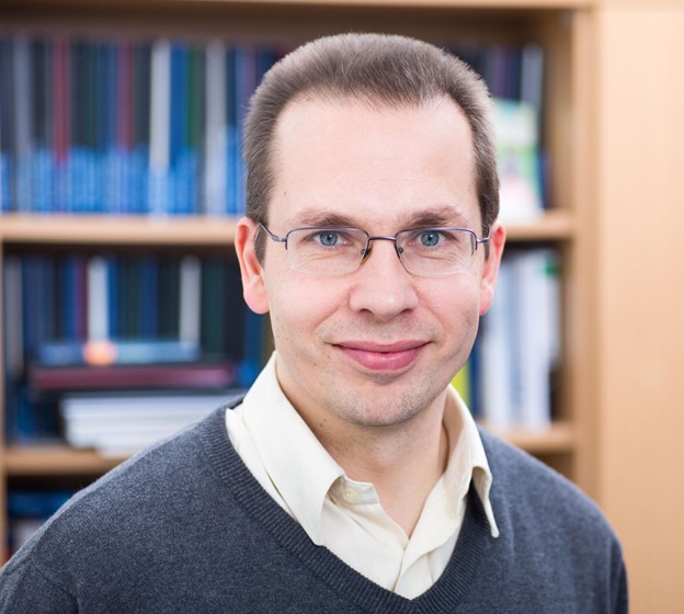
Bernhard Preim was born in 1969 in Magdeburg, Germany. He received the diploma in computer science in 1994 (minor in mathematics) and a Ph.D. in 1998 from the Otto-von-Guericke University of Magdeburg. In 1999 he moved to Bremen where he joined the staff of MeVis and directed the "computer-aided planning in liver surgery" group. In June 2002 he received the Habilitation degree (venia legendi) for computer science from the University of Bremen. Since Mars 2003 he is full professor for "Visualization" at the computer science department at the Otto-von-Guericke-University of Magdeburg, heading a research group focussed on medical visualization. He authored several textbooks: "Entwicklung interaktiver Systeme" (1999), "Visualization in Medicine" (Co-author Dirk Bartz, 2007), "Interaktive Systeme" (Co-author: R. Dachselt, 2010) and "Visual Computing in Medicine" (Co-author: C. Botha, 2013) and "Interaktive Systeme, Part 2" (Co-author: R. Dachselt, 2015) and "Visualization, Visual Analytics and Virtual Reality in Medicine" (Co-Authors: Renata Raidou, Noeska Smit and Kai Lawonn, 2023).
Bernhard Preim founded the working group Medical Visualization in the German Society for Computer Science and served as speaker from 2003-2012. He was president of the German Society for Computer- and Robot-Assisted Surgery (www.curac.org). He was Co-Chair and Co-Organizer of the first and second Eurographics Workshop on Visual Computing in Biology and Medicine (VCBM) and lead the steering committee of that workshop until 2019.
He is the chair of the scientific advisory board of ICCAS (International Competence Center on Computer-Assisted Surgery, since 2010), was member of advisory board of Fraunhofer Heinrich-Hertz-Institute (2008-2017) and member of the advisory board of Institute for Surgical Training and Technology Leipzig (2012-2018). From 2011-2018 he was an associate editor of IEEE Transactions on Medical Imaging and and IEEE Transactions on Visualization and Graphics (2017-2022). Currently he serves in the editorial board of Computers & Graphics (since 2019). He was paper co-chair of VMV 2012, EuroVis 2013 and Area Chair of IEEE VisWeek 2021.
He was also regularly a Visiting Professor at the University of Bremen where he closely collaborates with Fraunhofer MEVIS (2003-2012) and was Visiting Professor at TU Vienna (2016). At the University of Magdeburg, Bernhard Preim is member of the Board (since 2008), and was Vice-Dean for Student Affairs from 2012 to 2018). Bernhard Preim was married with Uta Preim (Medical Doctor), born Hahn (she died in 2021).
Abstract: In this "Ask-Me-Anything" session, Prof. Bernhard Preim will share his insights about his journey in academia, why he chose to stay in the field, and highlights from his research. Afterward, students will have the opportunity to engage in an interactive discussion, asking him anything---from his research expertise to advice on pursuing an academic career. This is a great chance to learn from an expert and get personalized guidance for your own academic path!
12:00 - 13:00
Lunch Break
13:00 - 14:00
Augmented Reality Visualizations for Interventional Navigation
Speaker: Florian Heinrich (Univ. Magdeburg)
Room: 335
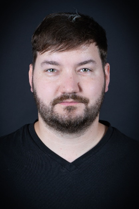
Dr. Florian Heinrich is currently a post-doc researcher at the Virtual and Augmented Reality Group at the Otto von Guericke University Magdeburg, Germany. He did his PhD studies on medical AR visualizations in the field of needle-based interventions. There, his main focus was on projector-based AR solutions. He has also previously worked as a senior researcher at the chair for Human-Computer Interaction of the University of Würzburg, Germany. He is passionate about novel AR and VR technologies and his primary research interests include visualization and interaction concepts for these devices. As part of the Research Campus STIMULATE, he is working on various medical application scenarios of AR and is interested in simulating medical workflows and projector-based visualizations in VR.
Abstract: In this presentation, we will discuss all relevant steps leading up to augmented reality (AR) visualizations for interventional navigation. This tutorial starts with fundamentals of interventional navigation to learn about relevant technical parts and terms. We will then talk about possible registration and tracking solutions, as well as AR devices to display our content. Following up some design considerations for AR navigation visualizations are presented. Finally, we will discuss current challenges and limitations in this field.
14:00 - 15:00
Uncertainty in Diffusion MRI Tractography
Speaker: Thomas Schultz (Univ. of Bonn)
Room: 335
Thomas Schultz is a professor at the University of Bonn, where he is heading the Visualization and Medical Image Analysis Group, and a PI at the Lamarr Institute for Machine Learning and Artificial Intelligence. He works on the development and integration of computational tools for quantitative image analysis, machine learning, and interactive visualization, in order to gain insights from large, complex, and dynamic image data. Prof. Schultz received his MSc (Dipl.-Inf.) and PhD (Dr.-Ing.) degrees in computer science from Saarland University and MPI Informatik in 2006 and 2009, respectively, after studying computer science and medical engineering in Saarbrücken, Heidelberg/Heilbronn, and Linköping (Sweden). He spent two years (2009-2011) as a DAAD postdoctoral fellow at the University of Chicago, and another two years (2011-2013) at the MPI for Intelligent Systems in Tübingen.
Abstract: Diffusion MRI tractography reconstructs white matter tracts in the human brain from noninvasive measurements, and is widely used in surgery planning and neuroscience. However, tractography is affected by various types of uncertainty, caused by measurement noise, loss of information through partial voluming, and the need for simplifying assumptions during data analysis. This talk will provide an introduction to diffusion MRI tractography, highlight some of its applications, and discuss approaches towards more reliable interpretation via uncertainty visualization and uncertainty reduction.
15:00 - 15:15
Coffee Break
Room: 301
15:15 - 16:15
Unlocking eXtended Realities for Medical Interfaces
Speaker: Daniel Simoes Lopes (Instituto Tecnico Superior)
Room: 335
Daniel Simões Lopes is a tenured Assistant Professor of the Computer Science & Engineering at Técnico Lisboa and VP of the Board of the Interactive Technologies Institute / LARSyS. He holds a degree in biomedical engineering from the University of Lisbon and graduated in computational engineering under the framework of the UT Austin|Portugal Program. At the educational level, he teaches Computer Graphics (undergraduate) and Virtual Reality (graduate) courses. At ITI, he directs the Lab of xReality and investigates novel XR interfaces with applications in 3D content creation and medical scenarios, as well as improved Computer Graphics techniques to better interact in the Metaverse.
Abstract: Extended Reality (XR) medical interfaces refer to interactive systems that use emerging technologies such as Virtual Reality (VR), Augmented Reality (AR), and Mixed Reality (MR) to advance the way medical care, training, and education are delivered and experienced. This talk will explore XR core concepts, unfold the design process, and provide guidelines for prototyping an XR medical interface. Additionally, several projects will be showcased, demonstrating the design of XR interfaces and interaction techniques for medical applications.
16:15 - 17:15
Visualization of 3D Spatial Biology
Speaker: Eric Mörth (Harvard)
Room: 335
Eric Mörth is a Post Doctoral Specialist Research Fellow in the Department of Biomedical Informatics at Harvard Medical School (DBMI) and part of the HIDIVE Lab. He received his PhD from the University of Bergen in Norway. During his PhD study, Eric Mörth conducted research in multimodal medical visualization. His focus was the research of new and innovative ways to visualize and explore medical data. Now Eric is researching the visualization of large-scale 3D Tissue Images using novel rendering techniques. Furthermore, his research enables the efficient analysis of highly multiplexed tissue images, and he also investigates how novel output devices like virtual and augmented reality devices can help investigators make better sense of their data.
Abstract: This lecture will explore the recent advancements in experimental techniques that allow biologists to capture the molecular properties of tissues in conjunction with their 3D spatial positioning within human organs. The focus will be on the generation and analysis of 3D Tissue Maps, which provide critical insights into the spatial organization and interactions of biological structures, from individual cells to complex tissue units. Despite the potential of these maps to enhance our understanding of healthy and diseased tissues, the field faces challenges in the analysis and visualization of this data. This lecture will address the importance of interpreting 3D Tissue Maps within their spatial context and discuss emerging strategies to improve their visualization and analysis.
17:15 - 17:30
Closing
Room: 335
8:00 - 9:00
Registration
Room: 218
9:00 - 10:00
Image-based blood flow simulation for risk assessment and virtual treatment of brain aneurysms
Speaker: Samuel Voß (Univ. Magdeburg)
Room: 335
Samuel Voß is currently a postdoctoral researcher at the Lab of Fluid Mechanics and Technical Flows. He holds a Ph.D. in Computational Fluid Dynamics, which he earned in 2022, following his studies in Mechanical Engineering. His current research is centered on the hemodynamic characterization of intracranial vessels and aneurysms, with the goal of improving risk assessment and virtual treatment. In previous projects, he has also contributed to the numerical simulation of airflow in the human larynx and the automatic characterization of ventral hernias using non-rigid CT image registration techniques.
Abstract: This presentation will introduce Computational Fluid Dynamics and explore its challenges when applied to clinical data. The focus will be on the simulation of hemodynamics inside intracranial vessels and aneurysms. A workflow analysis will highlight the steps involved from clinical imaging to risk scoring. Finally, the potential of this approach for improving risk assessment of diagnosed aneurysms and for simulating various treatment scenarios is discussed, offering new insights into patient-specific care and intervention planning.